 Healthcare · AI · Dental imaging
Skiavera
Open every scan already marked up.
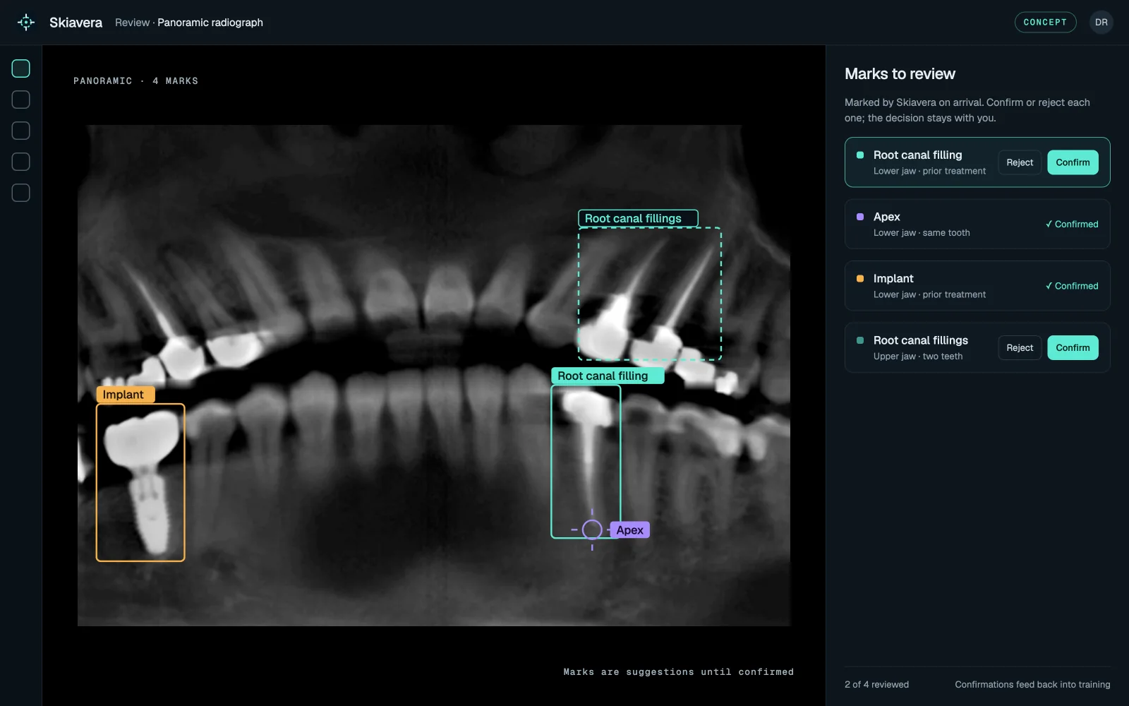
- 3 image typesPeriapical, panoramic and CBCT
- 1 expertTrained on one endodontist’s annotations
- You decideEvery annotation is confirmed or rejected by the clinician
Overview
Skiavera is an AI reader for dental radiographs. It reproduces one expert’s way of reading an image and applies it to every scan, so the clinician opens an image that is already marked up instead of a blank one.
Challenge
Reading a radiograph well takes years of experience, and two readers rarely mark the same image the same way. Finding every canal, apex, lesion and nearby structure takes time a busy practice doesn’t have, and any automation has to leave the diagnosis and the treatment decision with the clinician.
Solution
Skiavera is trained on periapical and panoramic X-rays and CBCT scans annotated by an experienced endodontist. It learns to find what that reader looks for: root canals with their number and curvature, the apex and periapical lesions, fractures, resorption, prior treatment, and anatomy that matters for planning, such as proximity to the sinus or the inferior alveolar nerve. When a new image arrives, those points are already marked on it. The clinician confirms or rejects each annotation, and those decisions feed back into training, so the model keeps learning from the practice that uses it.
Product
- 01Root canals, with their number and curvature
- 02Apex and periapical lesions
- 03Fractures, resorption and prior treatment
- 04Sinus and inferior alveolar nerve proximity for planning
- 05Confirm or reject every annotation
- 06Keeps learning from the practice’s own confirmations
Technology
- Computer vision
- Machine learning
System architecture
01Images
- Periapical
- Panoramic
- CBCT
02Model
- Annotation model
- Expert-labelled training set
03Review
- Clinician confirms or rejects
- Feedback into training
Outcome
The goal is consistency and speed, not replacement: every image is read the same careful way, and the clinician stays the decision maker. The first version is in development and being tested with a partner endodontic practice on its own images. Rootine Technology owns the model and the annotated dataset.